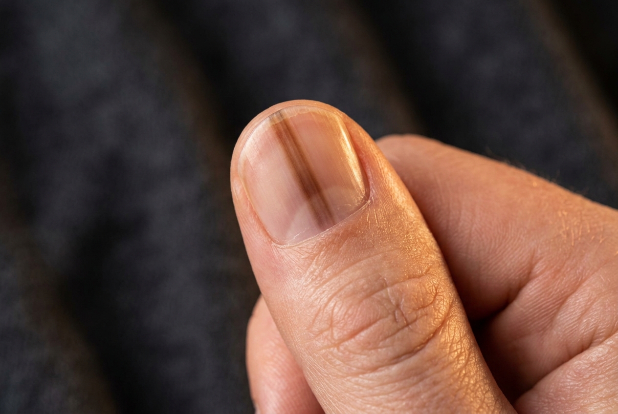
Atopic dermatitis on the face: how to recognize it and why it can leave dark spots?

Why I'm telling you this
There are patients who come to my office and tell me: 'Doctor, my face is dry, it itches, it turns red and flakes. Then it gets better, but I'm left with a dark spot for weeks or even months.' And another concern often arises: is the dermatitis still active? Have I got a scar? Why is my skin still dark if the eczema has already gone away?
I am Dr. Florian Vallecillo, and today I want to explain two aspects that are intimately connected: how to recognize atopic dermatitis on an adult's face, and why, after an inflammatory flare, the skin can remain darker for a long time.
Because inflammation is one thing. And the mark that inflammation can leave on the skin's pigmentation is quite another.
Watch the video
Atopic dermatitis: four simple answers
Can atopic dermatitis affect the face in adults?
Yes. Although many people associate atopic dermatitis mainly with children, it can also affect adults. And in adults, the face and neck can become particularly problematic areas: it can affect the eyelids, the area around the eyes, the forehead, the cheeks, the area around the mouth, and the neck. In some patients, dryness predominates; in others, redness; in others, itching; and some present a combination of all of these.
What can facial atopic dermatitis look like?
During a flare, we may find dry skin, scaling, redness, itching, a feeling of tightness, stinging, small cracks, inflamed plaques, and lesions caused by scratching. When inflammation persists for a long time and the patient scratches repeatedly, the skin can even thicken; we call this lichenification — the skin's response to the chronic cycle of itching, scratching, inflammation, and more itching.
But atopic dermatitis does not always appear red
This is a very important point. We are used to identifying inflammation with the color red, but that depends greatly on skin phototype. On lighter skin, active dermatitis may appear red or pink; on more pigmented skin, it may appear brown, violet, grayish, or simply darker than the surrounding skin, and sometimes the erythema is much less obvious. That is why we should not rely solely on the question 'Is it red?' to decide whether skin is inflamed: we also need to look at texture, scaling, edema, excoriations, itching, and clinical history.
Why is the face a particularly delicate area?
Because facial skin is not the same as the skin on the back or the legs. Especially around the eyes, the skin is much thinner. In addition, the face is constantly exposed to cosmetics, creams, makeup, perfumes, cleansers, sunscreens, hair products, pollution, cold, heat, sweat, and solar radiation. That is why, when persistent facial dermatitis appears, we should not automatically assume that 'it is just my usual atopic dermatitis.'
Not every facial eczema is atopic dermatitis
This is probably one of the most important messages in this article. Facial dermatitis can resemble many conditions: allergic contact dermatitis, irritant dermatitis, seborrheic dermatitis, rosacea, psoriasis, perioral dermatitis, and other inflammatory diseases. Furthermore, a person with atopic dermatitis has a more vulnerable skin barrier and can simultaneously develop contact dermatitis. In other words: having atopic dermatitis does not prevent someone from having another form of dermatitis at the same time.
A very common example: the eyelids
Eyelid dermatitis is extremely interesting. The patient thinks, 'I have atopic dermatitis around my eyes,' and that may well be the case; but the eyelids are also a very common site for contact dermatitis. And the responsible product does not even have to be applied directly to the eyelid: it can come from cosmetics, creams, perfumes, shampoos, hair products, nail polish, preservatives, fragrances, or substances transferred by the hands. That is why persistent or recurrent eyelid dermatitis may warrant a more specific investigation.
How do we tell one from the other?
There is no universal picture of 'facial atopic dermatitis.' The diagnosis depends on the appearance, distribution, itching, course, history of atopy, products used, response to previous treatments, and dermatological examination. In some patients, if we suspect a contact allergy, we can carry out patch tests, which attempt to identify substances capable of triggering a delayed allergic skin reaction.
How do we treat atopic dermatitis on the face?
First, by rebuilding the skin barrier. We use moisturising and emollient products suited to sensitive skin; we generally look for well-tolerated formulas, free of unnecessary fragrances, with few irritants, and appropriate for the facial area. But when active inflammation is present, moisturising alone may not be enough.
Can corticosteroids be used on the face?
Yes, but judiciously. Topical corticosteroids are extraordinarily useful for controlling a flare of dermatitis; however, the face — and especially the eyelids — requires more caution than other areas of the body. We do not necessarily use the same potency or for the same length of time as we would for a thick plaque on the hands or trunk. Treatment must be tailored to the location, severity, duration, and the individual patient.
There are also corticosteroid-free treatments
For certain facial dermatitis cases we use medications such as tacrolimus or pimecrolimus, which are topical calcineurin inhibitors. They have an important advantage in delicate areas: they do not cause the skin atrophy associated with prolonged corticosteroid use. That is why they can be particularly useful on the face, eyelids, neck, skin folds, and in maintenance strategies for certain patients.
Now let us imagine that we manage to bring the flare under control
The skin stops itching, the inflammation is gone, and the flaking disappears. But the patient comes back a few weeks later and tells me: 'Doctor, look. The dermatitis has gone, but I am left with this mark.' And this is where the second part of this story begins: post-inflammatory hyperpigmentation.
What is post-inflammatory hyperpigmentation?
The name sounds complicated, but the concept is very simple. Hyperpigmentation means that the skin produces or accumulates more pigment; post-inflammatory means that it occurs after inflammation. Therefore, post-inflammatory hyperpigmentation is a dark spot that appears after the skin has been inflamed. And it can occur after many situations: acne, insect bites, burns, dermatitis, trauma, psoriasis, skin procedures, and, of course, atopic dermatitis.
Why can inflammation change the color of the skin?
Here we need to talk about melanin, the pigment that determines a large part of our skin color and which is produced by cells called melanocytes. When inflammation is present, different molecules released in the skin can alter the activity of melanocytes, which may increase the production and transfer of melanin. The result is that the area where the eczema was can become darker. But there is another mechanism.
Sometimes the pigment can settle deeper
When the inflammation is significant enough, the basal layer of the epidermis can be damaged, and some of the melanin may then move into deeper layers of the skin. There it can be taken up by cells called melanophages. And when the pigment is located deeper, it can take much longer to disappear. This is why some spots fade relatively quickly while others can persist for months.
So, does the spot mean that the dermatitis is still active?
Not necessarily, and this is fundamental. A lesion can be free of itching, scaling, and active inflammation, and still appear darker. At that point we are seeing the consequence of the previous inflammation, not necessarily the current inflammation. This is why continually treating a residual spot as though it were still an active flare can be a mistake.
Is it a scar?
Generally, no. Post-inflammatory hyperpigmentation is an alteration in color, whereas a scar involves a deeper modification of the tissue's structure. A brown mark left after dermatitis does not automatically mean that the skin has permanently scarred; in many cases the color gradually lightens over time, though it can take a while.
Who is at greater risk of developing spots after dermatitis?
It can occur in any skin type, but it tends to be more intense, more noticeable, and more persistent in higher phototypes — that is, in people whose skin naturally produces more melanin. This is why a relatively brief episode of dermatitis can leave visible pigmentation for months in some people.
And why do some people end up lighter instead of darker?
That can also happen. After inflammation we may find either post-inflammatory hyperpigmentation or post-inflammatory hypopigmentation; in the latter, the area becomes temporarily lighter, something that is also seen in some forms of dermatitis. Therefore, after a flare the skin does not always immediately recover its original color.
Scratching makes the problem worse
Here we return to the infamous itch-scratch-inflammation cycle. The more we scratch, the more we traumatize the skin, the more we sustain the inflammation, the more we damage the barrier, and the greater the subsequent pigmentary alteration can be. This is why controlling the itch is not only about making the patient more comfortable: it also helps limit the consequences of inflammation.
And the sun can darken the patches even further
This is especially important on the face. Ultraviolet radiation stimulates melanogenesis — that is, it promotes the production of melanin. This means that an area already hyperpigmented after dermatitis can become even darker with sun exposure, and the contrast between normal skin and the pigmented area can also increase. Photoprotection is therefore a very important part of management.
Should I use sunscreen every day?
When facial hyperpigmentation is present, I do recommend adequate photoprotection, especially in places with high solar radiation such as Marbella. We look for a broad-spectrum sunscreen with a high SPF that is well tolerated by atopic skin. And that last point is essential, because there is no point recommending an excellent sunscreen if it irritates the skin and triggers another episode of dermatitis.
Visible light can also matter in certain types of pigmentation
Particularly in higher skin phototypes, visible light can contribute to some pigmentary alterations. For this reason, in certain patients with a significant tendency toward hyperpigmentation, tinted sunscreens that incorporate iron oxides may be of interest. However, this does not mean that every person with dermatitis necessarily needs a tinted sunscreen: photoprotection must be tailored to the individual patient.
How are patches treated after dermatitis?
Here the first rule is fundamental: control the dermatitis first. There is no point in attempting to depigment skin that remains inflamed, irritated, dry, itchy and with a compromised barrier, because some depigmenting treatments can irritate the skin; and if we irritate skin that already has a tendency to become inflamed, we can produce even more pigmentation.
First we put out the fire. Then we treat the scar it left behind
That is probably the simplest way to explain it. During the flare we control the inflammation; afterwards we repair the barrier; and once the skin is stable we assess whether we really need to treat the residual pigmentation. In many cases, simply time, photoprotection, control of new flares and good moisturisation allow for progressive improvement.
How long does it take for a patch to fade?
There is no universal timeframe. It depends on the intensity of the inflammation, the depth of the pigment, the phototype, sun exposure, the duration of the eczema, scratching, and whether or not new flares occur. Some patches improve within weeks, others require several months, and when the pigment lies deeper the recovery can be even slower.
Can depigmenting agents be used?
Yes, in certain patients — but skin with a history of atopic dermatitis requires particular caution. Depending on the case, different dermatological active ingredients aimed at modulating pigmentation may be used, but the priority is always to avoid re-inflaming the skin. Because a paradox emerges here: we use a product that is too aggressive to remove a blemish, the product irritates, the skin becomes inflamed, and the inflammation produces even more hyperpigmentation. That is why treatment must be gradual and adapted to cutaneous tolerance.
What about peels or lasers?
These can also have indications in certain types of hyperpigmentation, but not during active dermatitis. And we must be especially careful in patients with reactive skin, higher phototypes, or a tendency to hyperpigment, because an overly aggressive treatment can actually worsen the very problem we are trying to correct. That is why, before treating a blemish, we need to know what type of pigmentation it is, how deep it lies, what the phototype is, and whether the inflammatory condition is truly under control.
A blemish on the face does not always come from dermatitis
This point is also very important. A person with atopic dermatitis can simultaneously have melasma, solar lentigines, post-acne pigmentation, pigmented contact dermatitis, or other alterations. That is why we should not automatically assume: 'I have atopic skin, so all my blemishes are from the dermatitis.' In dermatology, we first diagnose the blemish and then decide how to treat it.
How can I prevent new blemishes from appearing?
The most effective strategy is to reduce repeated inflammation. That means properly maintaining the skin barrier, treating flare-ups early, avoiding irritating products, controlling itching, avoiding scratching, using photoprotection, and identifying any associated contact dermatitis. Because each new inflammatory episode can leave a new pigmentary mark.
A very common mistake: constantly switching cosmetics
When a patient sees dry skin, they buy a cream; then another one; then a serum; then an acid; then vitamin C; then retinol; then an exfoliant because they want to remove the blemish. And skin that already had a compromised barrier ends up receiving six or seven different products. The result is more irritation, more inflammation, more itching, and potentially more pigmentation. On atopic facial skin, less is very often more.
What basic routine might make sense?
It will depend on the patient, but conceptually we look for a gentle cleanser, adequate moisturisation, an anti-inflammatory treatment when indicated, photoprotection, and the avoidance of unnecessarily irritating products. Afterwards, once the skin is stable, we can decide whether we need to add a specific treatment for the pigmentation.
When should I seek advice?
When facial dermatitis persists, keeps coming back, affects the eyelids, causes significant itching, does not respond to standard treatment, or appears after introducing new cosmetics. Also when the patches change, are very persistent, do not clearly correspond to the areas of dermatitis, or we are simply not sure of the diagnosis. Because before attempting to depigment we need to know what we are treating.
What I want you to remember
Atopic dermatitis can affect the face in adults and cause dryness, flaking, inflammation, itching, burning, and subsequent pigmentation changes. But not every facial eczema is atopic dermatitis: especially when it affects the eyelids, face, and neck, we must also consider contact dermatitis and other dermatological diagnoses. And after a flare, even once the inflammation has cleared, the skin may remain dark. This is called post-inflammatory hyperpigmentation secondary to atopic dermatitis; it does not necessarily mean the disease is still active or that a scar has formed, but rather that the inflammation has temporarily altered the production and distribution of melanin.
I am Dr. Florian Vallecillo. And if I could leave you with just one idea, it is this: when the eczema disappears, the skin may need much more time to forget that it was inflamed. That is why the correct treatment does not consist solely of eliminating the patch; first we must control the inflammation, repair the barrier, prevent new flares, and protect the skin from the sun — and only afterwards, if the pigmentation persists, treat the pigment in a targeted way without re-inflaming the skin.
If you have atopic dermatitis on the face, recurrent eyelid eczema, or dark patches that persist after flares, a dermatological assessment at Clínica Valorian in Marbella makes it possible to distinguish atopic dermatitis from other causes of facial eczema and to determine whether the patches truly correspond to post-inflammatory hyperpigmentation. Because in atopic skin, getting the inflammation to resolve is the first step; getting the skin to then recover its balance, its barrier, and its colour may take a little more time.
Frequently asked questions about facial atopic dermatitis and dark patches
How do I know if I have atopic dermatitis on my face?
It can cause dryness, itching, flaking, inflammation, and a burning sensation, but there are other conditions with similar symptoms. Persistent facial dermatitis must be correctly diagnosed before it is treated.
Why does a brown patch remain after eczema?
Inflammation can stimulate melanocytes and alter the distribution of melanin. This is what we call post-inflammatory hyperpigmentation.
Does the patch mean I still have dermatitis?
Not necessarily. The inflammation may have resolved while the pigmentary change persists for weeks or months.
Are the patches permanent?
Usually not, although some may take months to fade. The duration depends on skin phototype, the intensity and depth of the inflammation, sun exposure, and the appearance of new flares.
Can I use retinol or acids to get rid of dark spots?
It is not a good idea to start potentially irritating treatments while the dermatitis is active. On atopic skin, triggering new inflammation can increase hyperpigmentation.
Does the sun darken spots?
Yes. Radiation can promote pigmentation and increase the contrast of residual lesions. Sun protection is especially important when facial hyperpigmentation is present.
Why do my dark spots last longer if I have dark skin?
Higher skin phototypes have a greater melanogenic capacity, and post-inflammatory hyperpigmentation can be more intense and persistent. This is why preventing new inflammatory episodes is particularly important.
What to remember
- —Atopic dermatitis also affects adults, and on the face and neck — eyelids, the area around the eyes, cheeks, and the perioral zone — it can be especially troublesome.
- —Inflammation does not always look red: in higher skin phototypes it can appear brown, purplish, or grayish. Texture, scaling, edema, itching, and clinical history must all be assessed — not just color.
- —Not every facial eczema is atopic dermatitis: it may be contact dermatitis (watch the eyelids!), seborrheic dermatitis, rosacea, psoriasis, or perioral dermatitis, and these conditions can coexist.
- —After a flare, post-inflammatory hyperpigmentation can remain: the inflammation alters melanocytes and melanin; if the pigment settles deep (melanophages), it takes longer to fade.
- —A residual spot does not mean active dermatitis or a permanent scar; it usually fades over time with flare control, good moisturization, and photoprotection.
- —First put out the fire, then address the mark: do not attempt depigmentation on inflamed skin (risk of further pigmentation); peels or laser only when the condition is under control; be cautious with an excess of cosmetic products.
You may also like · Skin & spots

Doctor Florian A. Vallecillo Cabrera
The doctor explains
Informational content, written and reviewed by Doctor Florian A. Vallecillo Cabrera. It does not replace an in-person consultation or an individual diagnosis.