Pityriasis versicolor and the other pityriasis conditions: understanding summer patches, avoiding confusion and preventing recurrence
Common in summer, pityriasis versicolor causes light, brown or pink patches on the chest, back and shoulders. Linked to a yeast normally present on the skin, it reflects neither poor hygiene nor an infection caught at the beach.

Doctor Florian A. Vallecillo Cabrera
Author
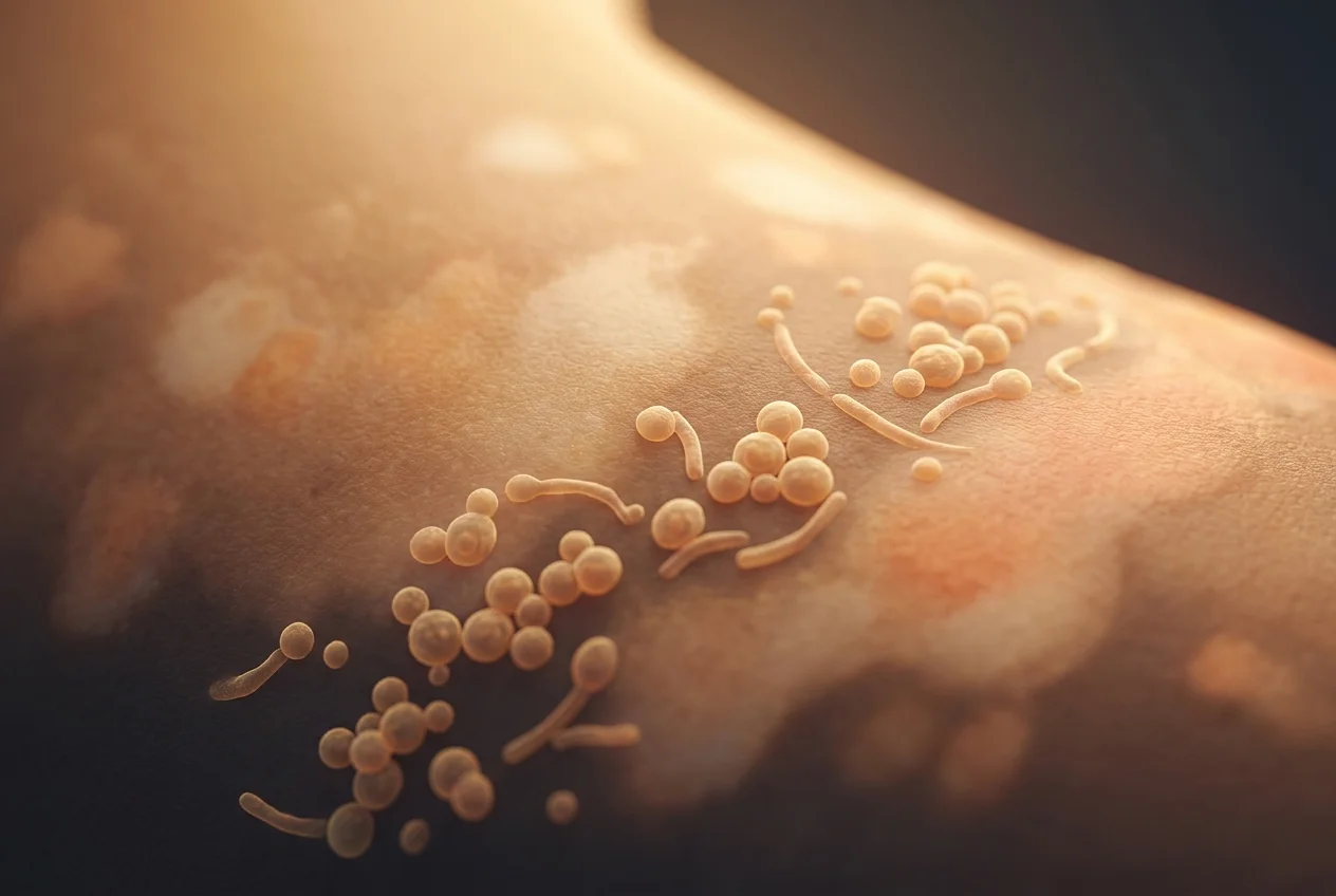
Pityriasis versicolor is a superficial fungal infection caused by the proliferation of Malassezia yeasts, which are normally present on the skin. It produces fine light, brown or pink patches on the trunk, especially visible in summer. Topical antifungals are first-line; oral ones are reserved for extensive or recurrent forms. Pigmentation can take weeks to months to normalise, and recurrences are frequent because the yeast is commensal. The term 'pityriasis' also covers very different diseases (alba, rosea of Gibert, rubra pilaris, lichenoides).
Summary
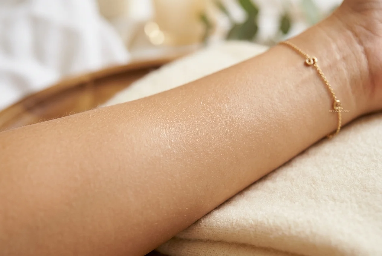
Pityriasis versicolor is a superficial skin condition caused by the proliferation of yeasts of the genus Malassezia, which normally belong to the skin microbiota. The lesions take the form of small light, brown, pink or coppery patches, covered with very fine scaling. They are located mainly on the chest, back, neck, shoulders and upper arms.
The condition becomes particularly visible in summer. Heat, humidity, sweating and sebum-rich skin promote the transformation of Malassezia towards a form associated with the lesions. Tanning then increases the contrast between the affected areas and the surrounding skin.
Pityriasis versicolor is usually neither dangerous, nor linked to poor hygiene, nor truly contagious in the usual sense of the term. Topical antifungals are the first-line treatment. Oral treatments are reserved for extensive, resistant or highly recurrent forms. Even after the yeast has been eliminated, pigmentation differences can persist for several weeks, sometimes several months.
The term "pityriasis", however, does not designate a single disease. Pityriasis alba, pityriasis rosea of Gibert, pityriasis rubra pilaris and pityriasis lichenoides have very different causes, mechanisms and treatments.
Why is pityriasis versicolor talked about so much in summer?
Pityriasis versicolor can be present all year round, but it is much more often noticed during the warm months. Several phenomena explain this:
- heat and humidity promote the growth of Malassezia;
- sweating creates a humid, occlusive skin environment;
- sebum provides the lipids these yeasts need;
- sports or very tight clothing increases occlusion;
- tanning makes the light patches more visible.
The sun is therefore generally not the direct cause of the lesions. It acts above all as a visual revealer: healthy skin tans, whereas the affected areas tan less uniformly.
This distinction is important. Many patients think they have "caught a fungal infection at the beach" because the patches appeared after the first sun exposures. In reality, the lesions may have already been present, but were barely visible on non-tanned skin.
The word "pityriasis" does not designate a single disease
The term pityriasis historically derives from a word evoking fine scaling comparable to bran. It is therefore mainly an old descriptive term, not a single diagnosis. The main diseases bearing this name include:
- pityriasis versicolor, linked to Malassezia;
- pityriasis alba, generally associated with mild dermatitis;
- pityriasis rosea of Gibert, an acute inflammatory eruption;
- pityriasis rubra pilaris, a rare inflammatory dermatosis;
- pityriasis lichenoides, a group of inflammatory papular dermatoses;
- the old term pityriasis capitis, sometimes used for dandruff.
These conditions do not necessarily have a biological link between them.
What is pityriasis versicolor?
Pityriasis versicolor, also called tinea versicolor in the English-language literature, is a superficial fungal infection of the stratum corneum of the epidermis. It is caused by lipophilic yeasts of the genus Malassezia. Among the species most often implicated are:
- Malassezia globosa;
- Malassezia furfur;
- Malassezia sympodialis;
- Malassezia restricta.
The exact distribution of species varies according to regions, populations and the methods used to identify them. A molecular study published in 2023 found M. globosa and M. restricta among the species frequently detected in pityriasis versicolor samples.
A more recent study carried out in Brazil identified six different species in samples from patients with pityriasis versicolor or seborrhoeic dermatitis, illustrating the diversity of the genus Malassezia. These results are interesting from a microbiological point of view, but they do not mean that it is necessary to identify the species in every patient in routine practice.
Malassezia: a yeast normally present on the skin
The presence of Malassezia is not abnormal. These yeasts are part of the skin microbiota of many healthy people. They live preferentially on regions rich in sebaceous glands:
- scalp;
- face;
- chest;
- back;
- shoulders.
The disease appears when certain conditions allow excessive proliferation or a change in the biological form of the yeast. Pityriasis versicolor can therefore be compared not to a classic external contamination, but to a local disruption of the balance of the skin microbiota. This distinction explains why:
- several members of a family may not be affected despite living together;
- the disease recurs after effective treatment;
- it is impossible and pointless to permanently eradicate all the yeasts on the skin.
What factors promote pityriasis versicolor?
The main associated factors include:
- hot and humid climate;
- heavy sweating;
- frequent sports activity;
- oily or seborrhoeic skin;
- adolescence and young adulthood;
- occlusive clothing;
- repeated application of very greasy products;
- individual predisposition;
- certain forms of immunosuppression;
- corticosteroid or immunosuppressive treatment in some contexts.
The majority of affected patients are, however, healthy. Pityriasis versicolor should therefore not automatically lead to a search for an immune disease. A broader evaluation is justified mainly in the face of very extensive, unusual, resistant forms or those accompanied by other infections.
Is it a contagious disease?
Pityriasis versicolor is generally not considered a highly contagious disease. As the responsible yeast is already present on the skin of a large part of the population, the development of lesions depends more on the skin terrain than on direct transmission between people. It is therefore usually not necessary to:
- isolate the patient;
- disinfect the whole house;
- systematically treat close contacts;
- throw away clothing;
- avoid the swimming pool for fear of contaminating others.
Usual hygiene, regular washing of clothes and correct drying of the skin are sufficient.
What does pityriasis versicolor look like?
The lesions generally consist of small round or oval macules that can merge to form larger plaques. Their colour varies:
- off-white;
- beige;
- light brown;
- café-au-lait;
- pink;
- salmon;
- coppery.
This diversity explains the term "versicolor". Very fine scaling is often present. It can be almost invisible at rest and appear when the lesion is gently scratched or the skin is stretched.
The most frequent areas are the upper back, the chest, the shoulders, the neck, the proximal part of the arms and the abdomen. In children, the face may be more affected than in adults. Itching is usually absent or mild, but it may be more marked in the case of heat or sweating.
Why do the patches turn white?
The light patches of pityriasis versicolor generally do not correspond to a definitive destruction of the melanocytes. Several mechanisms are proposed:
- transient disruption of melanin production or transfer;
- production of metabolites by Malassezia;
- modification of the stratum corneum;
- mild local inflammation;
- limitation of tanning in the affected areas.
The skin can thus remain lighter after the yeast has disappeared. This residual hypopigmentation is particularly visible in summer, because the surrounding skin tans more.
Why are some lesions brown or pink?
Pityriasis versicolor is not always white. The dark or pink forms may be linked to:
- post-inflammatory pigmentation;
- a local increase in melanin;
- a vascular reaction;
- the variable thickness of the stratum corneum;
- the patient's phototype.
In the same person, several colours can coexist.
How is the diagnosis confirmed?
Clinical examination
In a typical form, the diagnosis is often based on: the location; the variable colour of the lesions; their fine scaling; their tendency to confluence; their accentuation in hot climates.
The provoked scaling sign
Gentle scratching can bring out very fine powdery scaling. This sign is useful, but it is not perfectly specific.
Direct microscopic examination
A sample of scales can be examined after preparation with potassium hydroxide. The microscope classically shows clustered rounded yeasts and short mycelial filaments. This examination is particularly useful when there is doubt with vitiligo, pityriasis alba, seborrhoeic dermatitis, dermatophytosis or post-inflammatory hypopigmentation.
The Wood's lamp
Some lesions produce a yellow, yellowish-green or coppery fluorescence. However, the fluorescence is inconstant. A negative Wood's lamp therefore does not exclude the disease.
Dermoscopy
Dermoscopy can reveal fine scaling, scales located in the skin furrows, irregular pigmentation and sometimes jagged or follicular borders. Recent studies on hypopigmented dermatoses show that dermoscopy can help to differentiate pityriasis versicolor, pityriasis alba and vitiligo, while remaining a complementary examination that does not always replace microscopy.
Culture and PCR
Culturing Malassezia is difficult because these yeasts have particular lipid requirements. Molecular techniques allow a more precise identification of species. In the 2023 study, PCR detected Malassezia in a large part of the microscopy-positive samples, but its performance was not perfect. These methods are mainly useful in research or in atypical situations. They are not necessary in the majority of routine cases.
Which diseases can resemble pityriasis versicolor?
Vitiligo
Vitiligo usually produces a more marked depigmentation, often milky white, without scaling. The borders are often sharper and the hairs can sometimes turn white. The Wood's lamp generally strongly accentuates the lesions.
Pityriasis alba
Pityriasis alba produces poorly defined light plaques, mainly on the face of children and adolescents. It is considered a mild manifestation of an eczematous background and not a fungal infection. A small dermoscopic study found four frequent features: structureless white areas; fine scales; indistinct outlines; normally pigmented hairs.
Post-inflammatory hypopigmentation
Past inflammation can leave a light patch for several weeks or months: eczema, irritation, a bite, psoriasis, a burn or an aesthetic procedure.
Idiopathic guttate hypomelanosis
It causes small well-defined white spots, often on the arms and legs, particularly in adults who have had chronic sun exposure. It is neither infectious nor related to Malassezia.
Psoriasis
Some minimally inflammatory plaques can be confused with a fungal infection, particularly when they are located on the chest or the scalp.
Dermatophytosis
Dermatophyte infections more readily produce ring-shaped lesions, with an active peripheral border. They are caused by other fungi and can be more contagious.
Hypopigmented mycosis fungoides
This rare form of cutaneous lymphoma can resemble a chronic hypopigmented dermatosis. It should mainly be considered in the face of persistent, progressive, atypical plaques that are resistant to the usual treatments.
How is pityriasis versicolor treated?
Topical treatments: first line
Most patients can be treated locally. The products used include: azole antifungals; ciclopirox; selenium sulfide; zinc pyrithione depending on the available formulations; some non-azole local antifungals.
The choice depends on the extent of the lesions, their location, the available products, skin tolerance, age and previous treatments. Shampoos and lotions are often practical when the back, chest and shoulders are extensively affected. A cream may be suitable for more limited lesions.
The available reviews conclude that topical treatments are first-line, because they offer a good efficacy-safety ratio, few interactions and a generally limited cost.
Oral antifungals
An oral treatment can be considered when the affected surface is very large, when recurrences are frequent, when local application is difficult, when several correctly used local treatments have failed, or when the diagnosis has been confirmed.
Itraconazole and fluconazole are the agents most often studied in this context. However, their prescription must take into account drug interactions, hepatic risk, cardiac history for certain molecules, pregnancy and concomitant treatments.
Oral ketoconazole must not be used for pityriasis versicolor because of its unfavourable safety profile, particularly hepatic. Oral terbinafine is also poorly suited to this superficial infection, whereas some topical forms may be active.
Chemical peels
A case report published in 2025 reported an improvement after two 30% salicylic acid peels in a patient with extensive involvement and difficulties adhering to conventional treatments. This publication is interesting, but it is based on a single observation.
It does not demonstrate that the peel is superior to antifungals, that it prevents recurrences better, that it is suitable for all phototypes, or that it can be performed without risk of irritation or dyschromia. This method therefore remains experimental and is not a standard treatment.
Do the patches disappear immediately after treatment?
No. Two objectives must be distinguished: stopping the proliferation of the yeast and restoring homogeneous pigmentation. The first objective can be achieved quickly. The second often requires more time. The colour can take several weeks, sometimes two to three months, occasionally longer after old or very extensive involvement.
Persistence of the light patches therefore does not necessarily mean that the treatment has failed. The elements suggesting persistent activity are rather: appearance of new lesions; scaling still present; progressive extension; positive direct examination.
Multiplying courses of treatment simply because the pigmentation has not yet returned can cause irritation and adverse effects without speeding up recovery.
Why are recurrences so frequent?
Recurrences do not necessarily mean that the treatment was ineffective. They are explained by the fact that Malassezia belongs to the normal microbiota, that the seborrhoeic terrain persists, that the hot and humid climate reappears, that sweating and occlusion recur, and that some people are biologically more predisposed.
Recent work is also exploring the role of biofilms, of the lipases produced by the yeasts, of environmental tolerance mechanisms and of variations in antifungal sensitivity. A laboratory study published in 2026 showed that prolonged exposures to certain azoles could select cross-resistance profiles in Malassezia yeasts. These results are scientifically important, but they do not prove that all clinical relapses are due to resistance.
Another recent study observed differences in minimum inhibitory concentrations between species and antifungals. The susceptibility methods for Malassezia are, however, not yet as standardised as for other fungi. In practice, poor adherence, incomplete application, a new seasonal flare or residual pigmentation are often more likely than true resistance.
Can recurrences be prevented?
In patients presenting one or more flares each summer, intermittent prevention can be discussed. It may rely on the periodic use of a topical antifungal product during the warm months.
In some highly recurrent forms, intermittent oral prophylaxis has been studied. One trial reported better prevention of relapses with a monthly itraconazole protocol than with placebo, but such treatment requires a medical prescription and an evaluation of the benefit-risk ratio.
Prevention need not systematically be drug-based. Some simple measures can help:
- showering after heavy sweating;
- quickly changing damp clothes;
- favouring breathable textiles;
- limiting very greasy body products on the trunk;
- drying the folds and the upper body properly;
- starting the preventive protocol early in highly recurrent patients.
These measures reduce the factors promoting proliferation, but do not guarantee complete prevention.
Pityriasis alba: a common false fungal infection in children
Pityriasis alba mainly affects children and adolescents. It often appears as light, poorly defined, slightly dry plaques that are little or not itchy, located on the cheeks, chin or arms. It is associated with dry skin or an atopic background.
The sun makes it more visible by tanning the surrounding skin, which explains why it is frequently confused with pityriasis versicolor during the summer. Treatment is based mainly on moisturising, gentle care, photoprotection and a mild anti-inflammatory treatment if necessary. Antifungals are generally not useful.
Pityriasis rosea of Gibert: an acute inflammatory eruption
Pityriasis rosea of Gibert is very different from pityriasis versicolor. It often begins with a larger initial plaque, called the herald patch. A few days later, multiple oval lesions appear on the trunk, often oriented along the skin tension lines. The disease is generally self-limiting in two to twelve weeks.
A reactivation of the human herpesviruses HHV-6 or HHV-7 is suspected, but the exact mechanism remains debated. Authentic pityriasis rosea must be distinguished from a pityriasiform drug eruption. This difference is relevant because a drug reaction may require the identification and sometimes the discontinuation of the responsible drug.
Depending on the presentation, the differential diagnosis notably includes: secondary syphilis, dermatophytosis, guttate psoriasis, drug eruption and nummular eczema. The therapeutic data are limited. The available Cochrane review concludes that the studies are few, small and of low to moderate level of evidence. Most forms mainly require symptomatic treatment of the itching and monitoring.
Pityriasis rubra pilaris: a rare inflammatory disease
Pityriasis rubra pilaris is a much rarer disease. It can cause red-orange plaques, follicular papules, thickening of the palms and soles, large inflammatory areas, islands of healthy skin between the plaques and sometimes erythroderma. This disease requires specialised dermatological management.
Treatments may include retinoids, methotrexate, phototherapy in some cases, off-label targeted biologic treatments and, more recently, JAK inhibitors in a few observations. Recent publications highlight therapeutic progress, but the absence of large randomised trials limits the strength of the recommendations.
Pityriasis lichenoides
Pityriasis lichenoides is a group of rare inflammatory dermatoses. Chronic pityriasis lichenoides and the acute form, also called PLEVA, are mainly distinguished. The chronic form produces small scaly papules evolving in flares. The acute form can produce inflammatory, crusted, vesicular, sometimes necrotic lesions.
A skin biopsy is often useful to confirm the diagnosis. This condition is not caused by Malassezia and is not treated like pityriasis versicolor.
When to consult for light or brown patches?
A consultation is recommended when:
- the patches are completely white;
- they have no scaling;
- the hairs turn white;
- the lesions spread despite a correctly followed treatment;
- the diagnosis is uncertain;
- the skin is painful, inflammatory or oozing;
- the lesions concern a very young child;
- they recur several times a year;
- the patient is immunosuppressed;
- a loss of sensation is associated;
- the plaques persist for months without a clear diagnosis.
A medical evaluation helps avoid two frequent errors: treating vitiligo or eczema as a fungal infection; multiplying antifungals on already inactive residual pigmentation.
What the studies do not demonstrate
The available knowledge does not demonstrate that:
- pityriasis versicolor is caught at the beach;
- it results from poor hygiene;
- all the light patches of summer are fungal infections;
- any persistence of colour means the yeast is still active;
- recurrences are always due to antifungal resistance;
- identifying the Malassezia species systematically improves treatment;
- an antifungal susceptibility test is necessary in common forms;
- chemical peels are superior to antifungals;
- food supplements prevent relapses;
- all the "pityriasis" conditions have a common origin;
- oral treatments should be used first-line.
Practical implications
Pityriasis versicolor is generally benign, but it can be very visible and psychologically distressing. The most rational strategy is based on four steps: confirming that it is indeed pityriasis versicolor; treating the proliferation of the yeast with an approach adapted to the extent; explaining that the pigmentation may recover slowly; planning seasonal prevention in highly recurrent patients.
The success of the treatment should not be judged solely on the colour of the skin a few days after the course.
The Valorian analysis
Pityriasis versicolor is a disease that is simple in principle, but frequently misunderstood. The first misunderstanding comes from the word "fungal infection". The patient often imagines an external contamination, whereas Malassezia is a normal member of the skin microbiota. The disease appears when the balance between the yeast, sebum, temperature, humidity and the skin's response changes.
The second misunderstanding concerns the sun. It does not necessarily create the lesions: it mainly reveals a pre-existing pigmentary anomaly. The third concerns treatment. Eliminating the fungal proliferation does not immediately restore the colour. The persistence of light patches after a course of treatment therefore does not automatically justify restarting or intensifying antifungals.
Recent molecular research shows a significant diversity of Malassezia species and suggests the existence of adaptive resistance mechanisms. These data enrich our understanding of the disease, but they should not lead to turning every recurrence into a complex infection. In the majority of cases, relapses are explained by the persistence of the favourable terrain and the commensal nature of the yeast.
Finally, the confusion between the different "pityriasis" conditions remains a source of errors. Pityriasis alba is mainly eczematous, pityriasis rosea is an acute inflammatory eruption, and pityriasis rubra pilaris is a rare, potentially severe disease. Their common name is historical, not pathophysiological.
The modern objective is therefore not just to prescribe an antifungal. It is to make the correct diagnosis, avoid unnecessary treatments, prevent recurrences when relevant and reassure without trivialising atypical forms.
Key points
- ◆Pityriasis versicolor is a superficial fungal infection caused by a proliferation of Malassezia yeasts, normal members of the skin microbiota — not poor hygiene, not an infection caught at the beach.
- ◆It becomes visible mainly in summer: heat, humidity, sweating and sebum promote the yeast, and tanning accentuates the contrast of the patches (light, brown or pink) on the chest, back and shoulders.
- ◆Topical antifungals are the first-line treatment; oral ones (itraconazole, fluconazole) are reserved for extensive, resistant or highly recurrent forms (oral ketoconazole to be avoided).
- ◆After the yeast is eliminated, pigmentation can take several weeks to several months to become homogeneous again: the persistence of light patches does not mean a treatment failure.
- ◆The word 'pityriasis' covers distinct diseases (versicolor, alba, rosea of Gibert, rubra pilaris, lichenoides); it should also be distinguished from vitiligo to avoid unnecessary treatments.
Valorian level of evidence
- Link between Malassezia and pityriasis versicolor(5/5, high)
The role of Malassezia is established by clinical, microscopic, microbiological and molecular data.
- Efficacy of topical antifungals(4/5, moderate to high)
First-line in the majority of forms; the trials remain heterogeneous as to molecules, concentrations and durations.
- Efficacy of oral antifungals(4/5, moderate to high)
Itraconazole and fluconazole effective in selected forms, but their interaction and side-effect profile requires reasoned medical use.
- Drug prevention of recurrences(3/5, moderate)
Some intermittent strategies are effective, but the optimal protocols are not perfectly standardised.
- Clinical resistance to azoles(2/5, still limited)
Resistance mechanisms are demonstrated in the laboratory, but their real weight in common recurrences remains uncertain.
- Salicylic acid peel(1/5, very low)
The available data is based on a single case report; this option remains experimental.
- Role of probiotics and diets(0/5)
No nutritional or probiotic strategy can currently be recommended as a specific treatment or prevention.
References
- Leung AKC, Barankin B, Lam JM, et al. Tinea versicolor: an updated review. Drugs in Context. 2022;11:2022-9-2.
- Karray M, McKinney WP. Pityriasis Versicolor — A Narrative Review on the Diagnosis and Management. Life. 2023;13:2097.
- Gupta AK, Foley KA. Antifungal Treatment for Pityriasis Versicolor. Journal of Fungi. 2015;1:13-29.
- Eghtedarnejad E, Khajeh S, Zomorodian K, et al. Direct molecular analysis of Malassezia species from the clinical samples of patients with pityriasis versicolor. Current Medical Mycology. 2023;9:28-31.
- Diversity and Antifungal Susceptibility of Malassezia spp. Isolated From Brazilian Patients With Pityriasis Versicolor and Seborrheic Dermatitis. 2026.
- Soo YZ, Lee SM, Dawson TL Jr, Leong C. Azole-Driven Cross-Resistance and Transporter Gene Expression in Malassezia Yeasts. Microorganisms. 2026;14:1315.
- Wahab MA, Kamal SB, Shahin MR, et al. Efficacy of Itraconazole in the Prevention of Recurrence of Tinea Versicolor: A Three Year Follow Up. Mymensingh Medical Journal. 2020;29:351-356.
- Swerdlick SS, Krivda KR, Cox JA. Successful Treatment of Tinea Versicolor With Salicylic Acid 30% Peel. Federal Practitioner. 2025;42:270-273.
- Thomas IN, James JJ, Bala A, et al. Usage of Dermoscopy as an Effective Diagnostic Tool in Pityriasis Alba. Cureus. 2023;15:e40271.
- Villalon-Gomez JM. Interventions for pityriasis rosea. Cochrane systematic review, updated through 2018.
- Ciccarese G, Serviddio G, Drago F. Pityriasis rosea and pityriasis rosea-like eruption: The distinction is relevant for diagnostic and prognostic reasons. Journal of Family and Community Medicine. 2024;31:82-83.
- Sagut P, McIntyre ER, Elston DM. Pityriasis rubra pilaris. Journal of the American Academy of Dermatology. 2025;92:376-378.
- Menta N, Vidal SI, Stines A, Friedman A. Precision, Research, Progress: Updates in the Management of Pityriasis Rubra Pilaris. Journal of Drugs in Dermatology. 2025;24:833-834.
Frequently asked questions
Is pityriasis versicolor linked to poor hygiene?
No. It results from a proliferation of yeasts normally present on the skin and from a local predisposition.
Can you catch it at the swimming pool?
This is not the usual mechanism. The heat, humidity and sweating associated with the pool can, however, promote its visibility or reappearance.
Can you go to the beach with pityriasis versicolor?
Yes, but the difference in pigmentation may become more visible. Appropriate photoprotection is recommended.
Does the sun cure pityriasis versicolor?
No. Tanning can mask some dark forms or accentuate the light forms, but it does not eliminate the yeast.
Are the white patches permanent?
Most often no. The pigmentation gradually returns after the infection is controlled, sometimes over several weeks to several months.
Should the partner be treated?
Not usually, unless they themselves have lesions requiring a diagnosis.
Can an antifungal be used all year round?
In some highly recurrent cases an intermittent preventive treatment can be considered, but continuous, unsupervised use is generally not necessary.
Can probiotics or a sugar-free diet prevent recurrences?
There is currently no solid clinical evidence to recommend probiotics or a sugar-free diet to treat or prevent pityriasis versicolor.
Topics